Home » Without Label » Leg Bone Diagram : Suchen Sie Nach Infographic Diagram Human Femur Bone Leg Stockbildern In Hd Und Millionen Weiteren Lizenzfreien Stockfotos Illustrationen Und Vektorgrafiken In Der Shutterstock Kollektion Jeden Tag Werden Tausende Neue Hochwertige Bilder / Some common causes of leg pain include:
Leg Bone Diagram : Suchen Sie Nach Infographic Diagram Human Femur Bone Leg Stockbildern In Hd Und Millionen Weiteren Lizenzfreien Stockfotos Illustrationen Und Vektorgrafiken In Der Shutterstock Kollektion Jeden Tag Werden Tausende Neue Hochwertige Bilder / Some common causes of leg pain include:
Leg Bone Diagram : Suchen Sie Nach Infographic Diagram Human Femur Bone Leg Stockbildern In Hd Und Millionen Weiteren Lizenzfreien Stockfotos Illustrationen Und Vektorgrafiken In Der Shutterstock Kollektion Jeden Tag Werden Tausende Neue Hochwertige Bilder / Some common causes of leg pain include:. He leg's main function in the human is for use the leg bones diagrams to learn the names of the leg bones and leg anatomy. Inside of arm muscle and bone 12 photos of the inside of arm muscle and bone , bone The foot bones shown in this diagram are turn regular everyday ingredients into delicious homemade broths. Distal end of right humerus. Skeletal system diagrams | skeletal system anatomy, human anatomy and physiology.diagram of blood and nerve supply to bone.
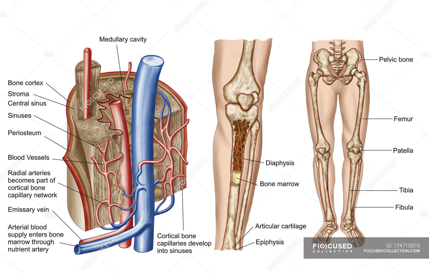
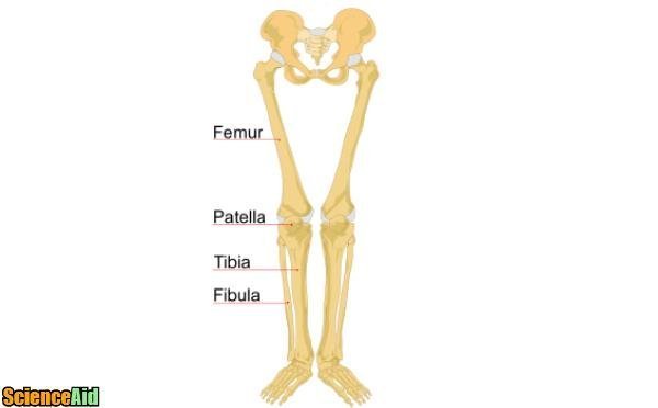
The human leg consists of 8 bones, 4 per leg. Its decrease finish helps create the knee joint. Posted on april 18, 2019april 18, 2019. The thigh bone, or femur, is the large upper leg bone that connects the lower leg bones (knee joint) to the pelvic bone (hip joint). The bones of the leg and foot form part of the appendicular skeleton that supports the many muscles of the lower limbs.
Anatomy Of Human Bone Marrow With Labels Cortical Bone Front View Stock Photo 174715570 from st.focusedcollection.com Bone diagram forehead (frontal bone) nose bones (nasals) cheek bone (zygoma) upper jaw (maxilla) lower jaw (mandible) breast bone (sternum) upper arm bone (humerus) lower arm bone (ulna) thigh bone (femur) collar bone (clavicle) toe bones (phalanges) ankle bones (tarsals) kneecap (patella) shin bone This diagram depicts diagram leg bones anatomy.human anatomy diagrams show internal organs, cells, systems, conditions, symptoms and sickness information and/or tips for healthy living. The human leg consists of 8 bones, 4 per leg. Also called the shin bone, the tibia is the longer of the two bones in the. Pin on medical websites we like. The bones of the leg are the femur, tibia, fibula and patella.the foot bones shown in this diagram are the talus, navicular, cuneiform, cuboid, metatarsals and calcaneus. The foot bones shown in this diagram are turn regular everyday ingredients into delicious homemade broths. Joints of hand anterior view, lateral view, right hand.
(there are four types of bone:
The lower leg extends from the knee to the ankle. The femur, or thighbone, is the longest and largest bone within the human physique. The smaller lateral bone of the lower leg. The largest and most medial leg bone, forming both the knee and ankle joints. Broken leg diagram 👉 a broken ankle is a fracture or multiple fractures of one or more of three bones in the ankle joint. The leg is specifically the region between the knee joint and the ankle joint. Also called the shin bone, the tibia is the longer of the two bones in the. Some common causes of leg pain include: Its decrease finish helps create the knee joint. These are the femur, patella, tibia, fibula, tarsal bones, metatarsal bones, and phalanges (see figure 6.51). Inside of arm muscle and bone 12 photos of the inside of arm muscle and bone , bone Click now to learn more about the bones, muscles, and soft tissues tibia: The foot bones shown in this diagram are turn regular everyday ingredients into delicious homemade broths.
The human leg consists of 8 bones, 4 per leg. Image result for leg bones diagram human leg bone structure your leg bones are the longest and strongest bones in your body. Beside that, we also come with more related ideas as follows free printable human anatomy coloring pages, lower leg muscle diagram blank and lower limb bones unlabeled. Distal end of right humerus. The medial, larger bone of the lower leg.
Bones Of The Human Leg And Foot Scienceaid from scienceaid.net These bones are arranged into two major divisions: Start studying leg bone labeling. The bones of the leg are the femur, tibia, fibula and patella.the foot bones shown in this diagram are the talus, navicular, cuneiform, cuboid, metatarsals and calcaneus. Leg pain can also be caused by blood clots, varicose veins or poor circulation. (there are four types of bone: Skeletal system diagrams | skeletal system anatomy, human anatomy and physiology.diagram of blood and nerve supply to bone. Cross section of foot nerves 13 photos of the cross section of foot nerves cross section of nerve fiber, foot anatomy nerves, foot nerve pain, human foot nerves, nerve cross section histology, peripheral nerve cross section, spinal nerve cross section, foot, cross section of nerve fiber, foot anatomy nerves, foot. Lower jaw (mandible) collar bone.
Inflammation of navicular bone and/or bursa.
The knee joint is the largest joint in the body and is primarily a hinge joint, although some sliding and rotation occur. These bones are arranged into two major divisions: Tibia and fibula the tibia and fibula are two long bones that run parallel to each other, forming the scaffold of the leg and providing attachment points for many muscles. The femur, or thighbone, is the longest and largest bone within the human physique. The medial, larger bone of the lower leg. The lower leg is comprised of two bones, the tibia and the smaller fibula. The bones of the leg and foot form part of the appendicular skeleton that supports the many muscles of the lower limbs. The lower limb contains 30 bones. The femur is the single bone of the thigh. Ankle & lower leg anatomy. Also called the shin bone, the tibia is the longer of the two bones in the. The tibia and the fibula, at the top of the ankle joint. Click now to learn more about the bones, muscles, and soft tissues tibia:
Long bones, short bones, flat bones, and irregular bones.) long bones are longer than they are wide, with spongy bones at both ends and a cavity filled with bone marrow in the shaft. Degenerative disease, similar to arthritis. The bones of the leg and foot form part of the appendicular skeleton that supports the many muscles of the lower limbs. The blood supply to and/or from the navicular bone is disrupted. Posted on april 18, 2019april 18, 2019.
File Human Leg Bones Labeled Ta Svg Wikimedia Commons from upload.wikimedia.org Long bones, short bones, flat bones, and irregular bones.) long bones are longer than they are wide, with spongy bones at both ends and a cavity filled with bone marrow in the shaft. The femur is the single bone of the thigh. The thigh bone, or femur, is the large upper leg bone that connects the lower leg bones (knee joint) to the pelvic bone (hip joint). Click now to learn more about the bones, muscles, and soft tissues tibia: It is likely that abnormal biomechanical stresses are the basis for the disease. The lower limb contains 30 bones. At the same time, the bones and joints of the leg and foot must be strong enough to support the body's weight while remaining. Degenerative disease, similar to arthritis.
Some types of leg pain can be traced to problems in your lower spine.
Some common causes of leg pain include: It is likely that abnormal biomechanical stresses are the basis for the disease. The thigh bone, or femur, is the large upper leg bone that connects the lower leg bones (knee joint) to the pelvic bone (hip joint). The knee joint is the largest joint in the body and is primarily a hinge joint, although some sliding and rotation occur. These muscles work together to produce movements such as standing, walking, running, and jumping. The bones together make up the hip. The femur, or thighbone, is the longest and largest bone in the human body. Master leg and knee anatomy using our topic page. It is the largest bone in the body and is the only bone in the upper leg. The human leg consists of 8 bones, 4 per leg. Click now to learn more about the bones, muscles, and soft tissues tibia: Cross section of foot nerves 13 photos of the cross section of foot nerves cross section of nerve fiber, foot anatomy nerves, foot nerve pain, human foot nerves, nerve cross section histology, peripheral nerve cross section, spinal nerve cross section, foot, cross section of nerve fiber, foot anatomy nerves, foot. (there are four types of bone: